Our Vision
Bridging Artificial Intelligence and Spatial Omics to Advance Precision Oncology
Motivation
Spatial biomarkers—defined as tissue-contextualized molecular, cellular, and architectural features that capture not only what is present but where and how biological entities are organized and interact within the tumor microenvironment—are central to enabling truly precise cancer diagnosis, prognosis, and treatment stratification.
Gap
Clinically established immunohistochemistry (IHC) workflows typically stain different markers on separate serial tissue sections, leading to loss of spatial continuity across markers and increased tissue consumption, which is particularly challenging with limited biopsy specimens. While state-of-the-art spatial omics technologies, including in situ multiplexed protein imaging and spatial transcriptomics profiling, can capture spatial biomarkers within the same tissue section, these approaches require specialized and costly instrumentation, reagents, and substantial wet-lab and analytical expertise, limiting their scalability and routine clinical adoption.
Our Digital Immune Reporter under the AI4HE Framework
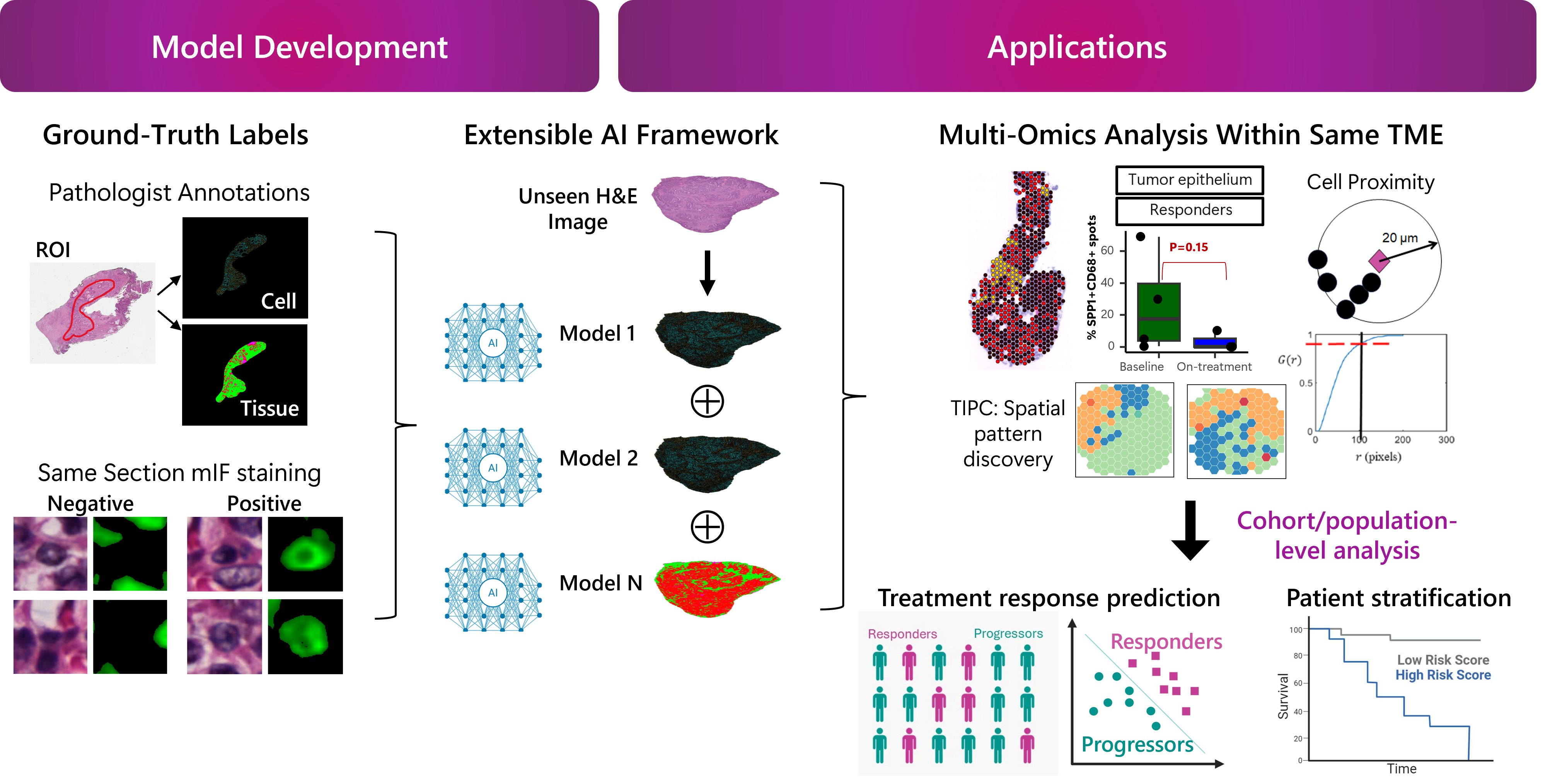
Building on the success of histology-based AI prediction, we extend this paradigm to uncover molecular and cellular information embedded within routine Hematoxylin and Eosin (H&E) images—an approach we term AI4HE.
This enables:
- Molecular-level insights without additional staining
- Single-cell-resolved inference within intact tissue architecture
- Scalable analysis across large clinical & public histology cohorts
- Seamless integration into existing digital pathology workflows
To further enhance interpretability and integration with established histopathological knowledge—such as pathologists’ grading and annotations used in routine clinical practice—we developed the Digital Immune Reporter (DIR): a one-stop, web-based platform for AI-driven prediction, spatial metric quantification, and interactive visualization of immune and tumor microenvironment features.